


- What Causes Glaucoma?
- Open Angle Glaucoma
- Closed-Angle (or Angle-Closure) Glaucoma
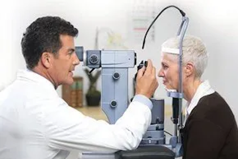
- Glaucoma Diagnostics
- Glaucoma Treatments
- Glaucoma Medications
- Glaucoma Laser
- Glaucoma Surgery
- Anatomic Narrow Angles
- Laser Peripheral Iridotomy
Glaucoma affects approximately 3 million Americans and over 120,000 will go blind from this disease. Glaucoma ranks as a leading cause of blindness worldwide. However, when glaucoma treatment starts early, vision loss from glaucoma is often preventable!
Glaucoma is a disease of the optic nerve, the large nerve that carries information from the eye to the brain. Glaucoma is caused by high pressure within the eye which results in damage to optic nerve fibers. The damaged nerve fibers cause blind spots or loss of vision, usually in the periphery of one’s vision. There are often no warning signs of glaucoma and patients are frequently unaware they have the disease until very late in the disease process. Once vision is lost, it cannot be regained. Early detection and treatment are key to preventing optic nerve damage and blindness from glaucoma.

What Causes Glaucoma?
Clear liquid, called the aqueous humor, circulates inside the front part of the eye. The eye is constantly producing a small amount of this fluid and an equal amount flows out of the eye through a microscopic drainage system. (This liquid is not part of the tears on the outer surface of the eye.)
The process of aqueous humor production and drainage is analogous to a bathtub with the drain open and faucet turned on. If the faucet is flowing faster than the water is draining from the tub, the tub will overflow. In the eye, if the rate of aqueous formation surpasses the rate of drainage, the pressure in the eye (the intraocular pressure) will increase. Chronic high pressure in the eye can result in progressive damage to the optic nerve head,
There are two major types of glaucoma, determined by the anatomy of the eye:
- Open-angle glaucoma
- Closed-angle glaucoma
Open Angle Glaucoma
Open angle glaucoma is the most common form of glaucoma in the United States. Pressure in the eye increases despite the presence of an open, or unobstructed, drainage system. With age, the drainage system of the eye becomes less efficient, resulting in decreased clearance of fluid. As the rate of fluid production outpaces the rate of outflow, pressure in the eye builds. This results in gradual, progressive, irreversible damage to the optic nerve.
There are very few symptoms associated with chronic open-angle glaucoma. There is no pain associated with this type of glaucoma and vision remains intact until late in the disease process. It is essential that patients are routinely screened for signs of glaucoma by an ophthalmologist.
Closed-Angle (or Angle-Closure) Glaucoma
In contrast to open angle glaucoma, angle-closure glaucoma is due to blockage of the eye’s drainage system. This blockage can be slow and gradual (over a period of years) or acute (over hours to days). A sudden, complete closure of the drainage apparatus of the eye causes a rapid increase in intraocular pressure and results in severe eye pain, headache (often severe enough to cause nausea and vomiting), blurred vision, glare and halos.
An ophthalmologist must evaluate each patient to determine the status of the angle and the function of the drainage system of the eye.
Glaucoma Diagnostics
Glaucoma can be detected and monitored by our ophthalmologist during routine eye examinations. Specific measurements and data are collected and analyzed, including:
- Tonometry– measurement of intraocular pressure
- Gonioscopy– assessment of the drainage angle of the eye
- Ophthalmoscopy– inspection of the optic nerve
- Optical Coherence Tomography– detailed measurements of the optic nerve thickness
- Perimetry– evaluation of peripheral vision
At Summit Eye Surgeons, all necessary testing can be completed in the comfort of the office.
Glaucoma Treatments
While damage caused by glaucoma cannot be reversed, there are several very effective methods to control intraocular pressure and slow the rate of glaucoma progression. These include eye drops, pills, laser procedures and surgical operations.
Glaucoma Medications
Glaucoma is frequently controlled with eye drops, sometimes in combination with oral or intravenous medications. These medications decrease eye pressure by either slowing the formation of aqueous fluids within the eye or by improving the flow through the drainage apparatus of the eye.
For these medications to work, they must take them regularly and continuously. Mild side effects from glaucoma medications are common and should be discussed with the ophthalmologist. The most frequent side effects include:
- Temporary stinging sensation
- Redness of the eye
- Changes in pulse or heart rate
- Difficulty breathing
- Headaches
- Blurred vision
Please contact Summit Eye Surgeons if you think you are experiencing a medication side effect before stopping the medication.
Glaucoma Laser
Selective laser trabeculoplasty (SLT) is a laser procedure used to lower intraocular pressure in patients with open angle glaucoma. Laser energy is applied to the trabecular meshwork (a specialized section of the draining system of the eye) to stimulate better flow through the eye’s drains. SLT is performed in an office-setting, requires very little pre-and post-procedural preparation. It is painless, safe and effective and repeatable.
A Peripheral Iridotomy is a procedure in which laser energy is used to make a tiny hole in the iris to enable fluid to escape from the front to the back of the eye if pressure were to increase. This procedure is only beneficial for patients with angle-closure or narrow-angle glaucoma.
Glaucoma Surgery
Glaucoma surgeries are reserved for more significant glaucoma cases in which the intraocular pressure cannot be adequately controlled through medications or lasers or in cases where a dramatic decrease in intraocular pressure is indicated. The two most commonly performed glaucoma surgeries are Minimally Invasive Glaucoma Surgery (MIGS), Trabeculectomy and Tube shunt.
Minimally Invasive Glaucoma Surgery (MIGS) refers to a group of surgical procedures that are generally faster, safer and have less side-effects than traditional glaucoma surgery. MIGS is generally reserved for mild to moderate cases of glaucoma and is performed in combination with cataract surgery, if there is a visually significant cataract.
A Trabeculectomy is a traditional glaucoma surgery in which an accessory drainage pathway is made for fluid to escape from the eye. A thin flap is made in the sclera (white outer wall of the eye) and a tunnel is created to help regulate flow of aqueous. The conjunctiva (thin, translucent tissue that overlies the sclera) is sutured closed to create a water-tight pocket called a bleb. The fluid that escapes from the eye is held in the bleb and then passes into the bloodstream.
A Tube shunt is a glaucoma draining implant that, like a trabeculectomy, facilities the drainage of fluid from the inside to regulate pressure in the eye. In this procedure, a tube (very small, straw-like devise) made of a synthetic material is placed in the front part of the eye and is tunneled to a pocket on the outer surface of the eye.
At Summit Eye partners, Dr. Lally can diagnose, monitor and treat all of your glaucoma needs to keep your eye pressures controlled and your optic nerve healthy.
Anatomic Narrow Angles
The angle of the eye is defined as the angle that is formed between the cornea (the clear, outer portion of the front part of the eye) and the iris (the colored part of the eye). The angle is where the draining apparatus of the eye is located. When angle is narrow, the drainage system is at risk for becoming so narrow that fluid can no longer escape. This could result in acute and chronic angle-closure glaucoma.
There are often no symptoms associated with anatomically narrow angles. While patients with anatomic narrow angles can have a predisposition to develop glaucoma if left untreated, they often do not require long-term glaucoma treatment. To prevent further narrowing of the angle and thus prevent acute and chronic angle closure glaucoma, a Laser Peripheral Iridotomy is needed.
Laser Peripheral Iridotomy
A Laser Peripheral Iridotomy is a procedure in which a laser is used to create a microscopic hole in the iris (the colored part of the eye) in patients with narrow or closed angles. This small hole acts as an escape route for fluid if the pressure in the eye should increase. This laser procedure is performed in an office-setting, requires very little pre-and post-procedural preparation/care. Rarely, laser peripheral iridotomy can cause temporary increased eye pressure and/or visual disturbances.
Related Blog Posts

Exploring Advanced Glaucoma Treatment Options: Minimally Invasive Solutions with Dr. Erin Lally